SpO2 vs. Pulse Oximetry: Choosing the Right Monitor
Pulse oximetry measures peripheral oxygen saturation. Dedicated pulse oximeters provide higher signal fidelity for critical care. Integrated SpO2 modules suit stable settings. Choose based on patient acuity, signal quality needs, and workflow requirements.
- Dedicated pulse oximeters deliver superior signal quality for unstable patients.
- Integrated SpO2 modules save space but may struggle with poor perfusion.
- Match the monitoring method to patient acuity and workflow constraints.
- Verify signal quality indicators before trusting saturation readings.
- Choose based on clinical setting rather than raw accuracy alone.
What Is the Core Difference?
Pulse oximetry refers to the optical technique for estimating arterial oxygen saturation. SpO2 is the specific measurement derived from that technique. In practice, the distinction matters less than the hardware delivering the signal. A dedicated pulse oximeter uses dedicated sensors, processing, and display logic. An integrated SpO2 module embeds the same photometry into a larger vital signs monitor.
The choice affects signal reliability, workflow, and maintenance. In stable patients, both methods often return similar saturation values. In hypotensive patients, those values can diverge significantly.
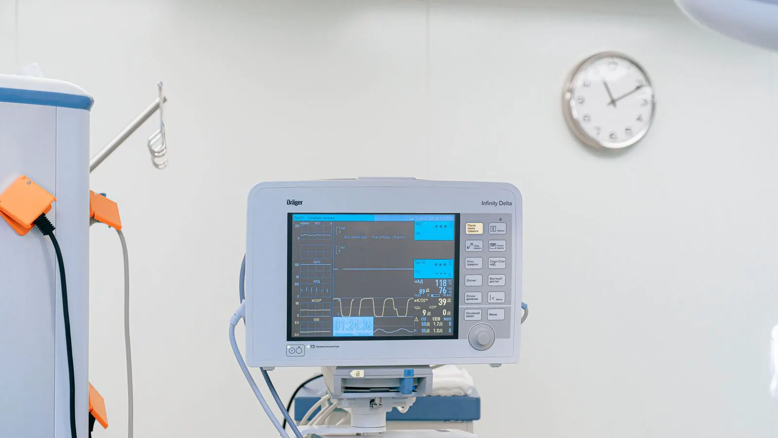
A dedicated pulse oximeter is a standalone unit. It connects to a single sensor, usually a finger clip, and processes the optical signal on its own internal hardware. The display shows only the saturation percentage, the pulse rate, and often a waveform trace. This isolation allows the manufacturer to optimize the entire processing chain for one variable. The device can sample the signal at high resolution and apply aggressive filtering without competing for processor cycles with other parameters.
An integrated SpO2 module is a board or sub-assembly inside a multiparameter patient monitor. It shares the same power supply, microcontroller, and display interface as the other channels. When the monitor is displaying ECG, blood pressure, and temperature, the SpO2 channel operates within a shared resource pool. The module receives the raw optical data, but the processing pipeline may be prioritized to maintain latency for critical waveforms or to manage power consumption.
This architectural difference creates a practical gap in performance. In a stable ward patient with good peripheral perfusion, the difference is often negligible. The saturation value on the monitor screen matches the reading on a standalone device. However, when perfusion drops, the shared processing may struggle to extract a clean signal from the noise. The dedicated device, with its dedicated resources, can often maintain a reading when the integrated module loses the pulse waveform.
How Do Accuracy Profiles Differ?
Accuracy depends on perfusion, sensor placement, and signal processing. Dedicated pulse oximeters typically use higher quality optical sensors and more sophisticated filtering algorithms. They handle motion artifacts and low perfusion better. This matters in intensive care units where patients may have vasopressor support or edema.
Integrated SpO2 modules on general patient monitors prioritize space efficiency. They often share processing resources with other parameters. This can introduce latency or noise. The result is acceptable for ambulatory or post-operative monitoring. It becomes problematic when small saturation drops indicate clinical deterioration.
Both systems rely on the same physics. They measure light absorption at two wavelengths. The difference is engineering execution.
The physics involves two infrared light sources, typically red at 660 nanometers and infrared at 940 nanimeters. Oxygenated hemoglobin absorbs red light more strongly than deoxygenated hemoglobin. The device measures the ratio of light transmitted through the tissue at these two wavelengths. This ratio is converted into a saturation percentage.
Dedicated devices often use more sensitive photodiodes. They can detect tiny changes in light absorption even when the blood volume pulsing in the capillary bed is low. The processing algorithms in these devices are tuned to distinguish the pulsatile arterial signal from non-pulsatile tissue absorption. They use adaptive filters that adjust in real-time based on the signal-to-noise ratio.
Integrated modules face a different constraint. The monitor must display multiple parameters simultaneously. If the SpO2 channel shares a processor with the ECG or blood pressure module, the SpO2 data may be sampled less frequently or with less computational power. This can result in a smoother but less responsive waveform. The saturation value may update more slowly, or it may drift slightly during periods of high motion.
In critical care, these small delays can matter. A rapid drop in oxygenation might be a sign of a developing complication. A dedicated device with a clear waveform and immediate saturation update allows the clinician to respond faster. An integrated module might show the same value but with a less clear trend, requiring the clinician to verify the reading with another method.
Comparison of Monitoring Approaches
| Option | Best for | Limitations |
|---|---|---|
| Dedicated Pulse Oximeter | Critical care, low perfusion states | Requires separate display, higher cost per unit |
| Integrated SpO2 Module | General wards, stable patients | May struggle with motion artifacts |
| Wearable SpO2 Patch | Long-term monitoring, ambulatory care | Limited to specific body sites |
| Finger Clip Sensor | Point-of-care checks | Difficult to use on patients with nail issues |
| Bedside Monitor Bundle | Multi-parameter monitoring | Signal quality varies by manufacturer |
When to Choose a Dedicated Pulse Oximeter
Select a dedicated pulse oximeter when the patient population includes unstable individuals. This applies to operating rooms, intensive care units, and emergency departments. These settings demand reliable saturation tracking during resuscitation.
Dedicated devices often display signal quality indicators. They show waveform clarity and perfusion strength. Clinicians can verify data quality before acting. This reduces false alarms and missed trends.
Maintenance is simpler. The sensor is a single point of failure. Replacement is straightforward. Calibration checks are quick. The device focuses on one function, which simplifies troubleshooting.
Cost per unit is higher. But the value comes from reduced false alarms and better clinical confidence. In high-acuity settings, that trade-off pays for itself.
In the operating room, a dedicated pulse oximeter is often attached directly to the patient’s hand or foot. The display sits on the anesthesia machine or a nearby cart. The anesthesiologist can watch the waveform while adjusting ventilation or administering medications. If the waveform flattens, the anesthesiologist knows immediately that perfusion has dropped. They can check blood pressure or reposition the sensor without waiting for a central monitor to update.
In intensive care units, patients may have multiple lines and tubes. A dedicated pulse oximeter can be placed at the bedside. The nurse can check the saturation quickly without moving to a central station. The device’s simple interface allows for fast verification of the reading. If the sensor is loose or the patient has moved, the waveform will show it clearly.
These devices also handle cold extremities better. In resuscitation, patients may have low core temperatures. Peripheral vasoconstriction reduces blood flow to the fingers. A dedicated pulse oximeter with advanced signal processing can often detect a pulse in this state. An integrated module might show no signal or an erratic value, leading the clinician to assume the sensor is faulty rather than the patient is perfusing poorly.
The cost of a dedicated unit is higher than the cost of an SpO2 channel built into a general monitor. However, the cost of a missed trend or a false alarm can be higher. In a high-acuity environment, the reliability of the dedicated device reduces the cognitive load on the clinical team. They can trust the reading and focus on patient care rather than questioning the equipment.
When to Choose an Integrated SpO2 Module
Integrated modules suit general wards and stable patient populations. They save desk space and reduce cable clutter. The patient monitor already displays blood pressure, heart rate, and respiration. Adding SpO2 to the same screen simplify workflow.
These modules work well for post-operative patients who are hemodynamically stable. They are suitable for telemetry setups where multiple parameters need display on one unit.
The limitation is signal fidelity. If the patient has poor peripheral circulation, the module may show a flatline or drifting values. Clinicians need to interpret this context. A dedicated pulse oximeter would likely provide a clearer signal in the same scenario.
Maintenance is more complex. The module is part of a larger system. Software updates may affect SpO2 processing. Sensor compatibility varies between monitor models.
In a general ward, the patient is usually stable. The room is quiet, and the patient is not undergoing resuscitation. An integrated module on a bedside monitor provides sufficient data for routine checks. The nurse can see the saturation value alongside the heart rate and blood pressure. If the saturation drops below a threshold, the monitor alarms. The nurse investigates.
The workflow benefit is significant. With a dedicated pulse oximeter, the nurse might need to check a separate screen or move the device. With an integrated module, all data is on one screen. This reduces the time spent collecting data and allows for faster response to changes.
However, the integrated module is not suitable for every situation. If a patient on the ward develops sepsis and their blood pressure drops, the SpO2 signal may degrade. The integrated module might struggle to maintain a clear reading. In this case, the clinical team might need to add a dedicated pulse oximeter to confirm the reading or switch to a different sensor site.
The maintenance aspect is also different. A dedicated pulse oximeter has a simple sensor that can be replaced in seconds. An integrated module is part of a larger monitor. If the SpO2 sensor fails, the entire monitor may need service, or the sensor may need to be replaced with a specific part that is only available from the manufacturer. This can cause downtime if the monitor is not available for replacement.
Key Selection Criteria for Clinical Use
Evaluate the patient population first. What is the typical acuity level? How often will you encounter low perfusion states? This determines whether signal quality is a primary concern.
Next, review the workflow. How many monitoring points does the room need? If the team already uses a central patient monitor, adding a dedicated pulse oximeter may create redundancy. If the patient requires frequent saturation checks, a dedicated device near the bedside improves accessibility.
Consider sensor handling. Dedicated pulse oximeters use standard finger clips. Integrated modules may use proprietary sensors or adhesive patches. Staff need training on correct placement. Improper placement causes false readings in any system.
Check signal quality display. Look for waveform visualization and perfusion indices. These features help clinicians distinguish real trends from noise. Without them, saturation values become less trustworthy.
Review maintenance requirements. Dedicated devices have replaceable sensors. Integrated modules require service contracts. Factor long-term cost into the decision.
Patient acuity is the primary driver. If the ward accepts patients with heart failure who are often on diuretics and may have low perfusion, a dedicated device might be necessary. If the ward accepts stable patients recovering from minor surgeries, an integrated module is sufficient.
Workflow considerations include the number of patients per nurse. If a nurse cares for four patients, each with a central monitor, the integrated module reduces cable clutter. If a nurse cares for two patients, one of whom is unstable, a dedicated pulse oximeter for the unstable patient provides a clearer signal.
Sensor handling is a practical issue. Standard finger clips are familiar to most staff. If an integrated module uses a proprietary sensor, staff need training to apply it correctly. If the sensor is an adhesive patch, it may be uncomfortable for long-term use. The choice of sensor affects patient comfort and data quality.
Signal quality display is a must-have feature. A saturation value without a waveform is less useful. The waveform shows the shape of the arterial pulse. A clear waveform indicates good perfusion. A flat or noisy waveform indicates poor signal. Clinicians need to see this to trust the reading.
Maintenance requirements affect long-term cost. A dedicated pulse oximeter has a sensor that costs a few dollars and lasts a few weeks. An integrated module may require a service contract that costs hundreds of dollars per year. Over five years, the cost of maintenance can exceed the cost of the hardware.
Common Misconceptions in Device Selection
Many buyers assume that higher accuracy always means better clinical outcomes. This is not always true. A highly accurate device that provides confusing alarms creates more problems than a reliable one with clear signals.
Some assume that SpO2 and pulse oximetry are interchangeable terms. They are not. Pulse oximetry is the method. SpO2 is the measurement. This distinction matters when evaluating device specifications.
Another common error is ignoring sensor placement. A perfect pulse oximeter gives wrong readings if the sensor is on a cold extremity. Training and protocol matter as much as hardware.
Finally, buyers often overlook the alarm management system. A good pulse oximeter with poor alarm configuration creates alert fatigue. Pair the device with appropriate alarm thresholds and escalation pathways.
The idea that higher accuracy always leads to better outcomes is a common trap. A device that is too sensitive may alarm for every small fluctuation. This creates alert fatigue, where staff ignore alarms because they are too frequent. A device that is slightly less accurate but provides a stable reading and clear alarms is more useful in practice.
The distinction between pulse oximetry and SpO2 is often blurred in marketing materials. Pulse oximetry is the process of measuring light absorption to estimate saturation. SpO2 is the specific value of oxygen saturation in the arterial blood. When buying a device, look at the specifications for the pulse oximetry method. Check the wavelengths used, the signal processing algorithm, and the display features. Do not just look at the SpO2 value.
Sensor placement is a critical factor. A pulse oximeter is only as good as the sensor placement. If the sensor is on a cold, numb finger, the reading will be inaccurate. Staff must be trained to place the sensor on a warm, well-perfused finger. They must also be trained to check the waveform to ensure the sensor is making good contact with the tissue.
Alarm management is often overlooked. A pulse oximeter that beeps constantly for small drops in saturation will be ignored by staff. The alarm thresholds must be set based on the patient’s condition and the clinical context. Escalation pathways must be defined. If the saturation drops below a certain level, who is notified? What action is taken? These protocols are as important as the device itself.
Implementation and Validation Checklist
Before deploying either system, validate performance in your specific environment. Run test cases with known saturation levels. Verify that the device tracks expected changes.
Train clinical staff on sensor placement and signal interpretation. Show them how to read waveform quality indicators. Discuss when to distrust the reading.
Document the monitoring protocol. Specify which patients receive dedicated pulse oximetry versus integrated SpO2 monitoring. Define escalation criteria for low saturation or poor signal.
Review the data output. Ensure saturation values integrate with your electronic health record. Check that trends display correctly over time.
Reassess performance annually. Sensor technology and clinical needs evolve. What works today may not fit tomorrow’s patient population.
Validation is a practical step. Before putting a new device into clinical use, test it in a controlled environment. Use a phantom or a volunteer with known saturation levels. Verify that the device reads correctly at different saturation levels. Check how the device behaves when the sensor is loose or when there is motion. This helps identify any issues before they affect patients.
Training is essential. Clinical staff need to understand how the device works. They need to know how to place the sensor correctly. They need to know how to interpret the waveform. They need to know when to distrust the reading. For example, if the waveform is flat but the patient is stable, the sensor may be mispositioned. If the waveform is clear but the saturation is low, the patient may be hypoxemic.
Documentation is a legal and practical requirement. The monitoring protocol should specify which patients receive which type of monitoring. It should define when to escalate for low saturation or poor signal. It should also specify how to handle sensor failures. This documentation protects the clinical team and ensures consistent care.
Data output is a growing concern. Saturation data should integrate with the electronic health record. This allows for long-term trends and easy access during handovers. Check that the data displays correctly over time. If the trend graph shows gaps or errors, the integration may need adjustment.
Annual reassessment is important. Sensor technology and clinical needs evolve. New sensors may offer better performance. Clinical needs may change as patient populations shift. What works today may not fit tomorrow. Regular review ensures that the monitoring system remains effective and efficient.
Final Selection Guidance
Choose a dedicated pulse oximeter when clinical acuity is high. This applies to critical care, operating rooms, and emergency settings. The investment in signal quality and reliability justifies the cost.
Choose an integrated SpO2 module for stable patients in general wards. The space savings and workflow simplicity outweigh the lower signal fidelity in most scenarios.
For ambulatory monitoring, consider wearable SpO2 patches. They extend monitoring beyond the hospital setting. Use them for patients who need long-term saturation tracking without continuous cable attachment.
Match the device to the clinical context. The right monitor is not the most accurate one. It is the one that delivers trustworthy data for the specific patient population and workflow.
The decision between a dedicated pulse oximeter and an integrated SpO2 module is not about which one is better. It is about which one fits the clinical context. A dedicated device is better for unstable patients. An integrated module is better for stable patients. A wearable patch is better for ambulatory monitoring.
The right monitor is the one that delivers trustworthy data for the specific patient population and workflow. It is the one that integrates well with the clinical team’s practices. It is the one that provides clear, reliable data without causing unnecessary alarms or confusion.
By matching the device to the clinical context, you ensure that the monitoring system supports clinical decision-making. This approach leads to better patient outcomes and a more efficient clinical workflow. It also reduces the risk of errors and missed trends. The goal is to provide the right data at the right time to the right clinician.
Frequently asked questions
Can I use a finger clip pulse oximeter in place of a patient monitor SpO2 module?
Yes, if the patient is stable and the clip is placed correctly. For unstable patients with low perfusion, a dedicated device provides more reliable signal.
How often should I replace pulse oximeter sensors?
Replace them when the adhesive fails or the window becomes dirty. Check during each patient change. Follow your facility's infection control protocol.
What causes flatline readings on an integrated SpO2 module?
Poor perfusion, sensor movement, or incorrect placement. Verify sensor position and check the patient's peripheral circulation.
Do dedicated pulse oximeters require separate calibration?
Most use factory calibration with periodic verification. Check your facility's policy. Some models allow field verification with test simulators.
How do I reduce false alarms from pulse oximetry?
Use proper sensor placement and adjust alarm thresholds. Avoid over-alarming. Train staff to interpret signal quality before acting on alerts.